Imaging tests let doctors look inside your body without surgery. Whether you have been told you need an X-ray after a fall, a CT scan for abdominal pain, an MRI for a knee injury, or an ultrasound during pregnancy, it helps to know what each test does, how safe it is, and what you need to do beforehand.
In this detailed guide, the Alaikas.com team explains radiology and imaging tests in plain language to make them less confusing, and it is part of our health topics hub. Below, you will find a clear comparison of the four most common imaging tests, what radiation risk really means, and practical steps to prepare for your appointment.
What Radiology and Medical Imaging Do
Radiology is the medical specialty that uses imaging to diagnose and sometimes treat disease. Two professionals are usually involved in your scan:
- A radiologic technologist (or sonographer, for ultrasound) positions you, operates the equipment, and makes sure the images are clear.
- A radiologist is a physician who interprets the images and writes a report for the clinician who ordered the test.
Imaging helps find broken bones, infections, tumors, blood clots, internal bleeding, and organ problems. It is also used to guide procedures such as biopsies and to track how a condition responds to treatment.
The Main Imaging Tests at a Glance
| Test | How it works | Often used for | Uses ionizing radiation? |
|---|---|---|---|
| X-ray | A small dose of radiation passes through the body to create a flat image | Broken bones, chest infections, dental problems | Yes, usually a low dose |
| CT scan | Many X-ray images taken from different angles are combined by a computer into cross-sections | Injuries, internal bleeding, lung and abdominal problems, cancer staging | Yes, higher than a standard X-ray |
| MRI | Strong magnets and radio waves create detailed images | Brain, spinal cord, joints, ligaments, soft tissues | No |
| Ultrasound | High-frequency sound waves bounce off tissues to create live images | Pregnancy, gallbladder, thyroid, blood flow, many abdominal organs | No |
How Each Imaging Test Works
X-rays
X-rays are the quickest and most widely available imaging test. You stand, sit, or lie still while the technologist takes one or more images, usually in a few minutes. Dense structures like bone appear white, while air in the lungs looks dark. The FDA explains the benefits and risks of medical X-ray imaging in more detail.
CT scans
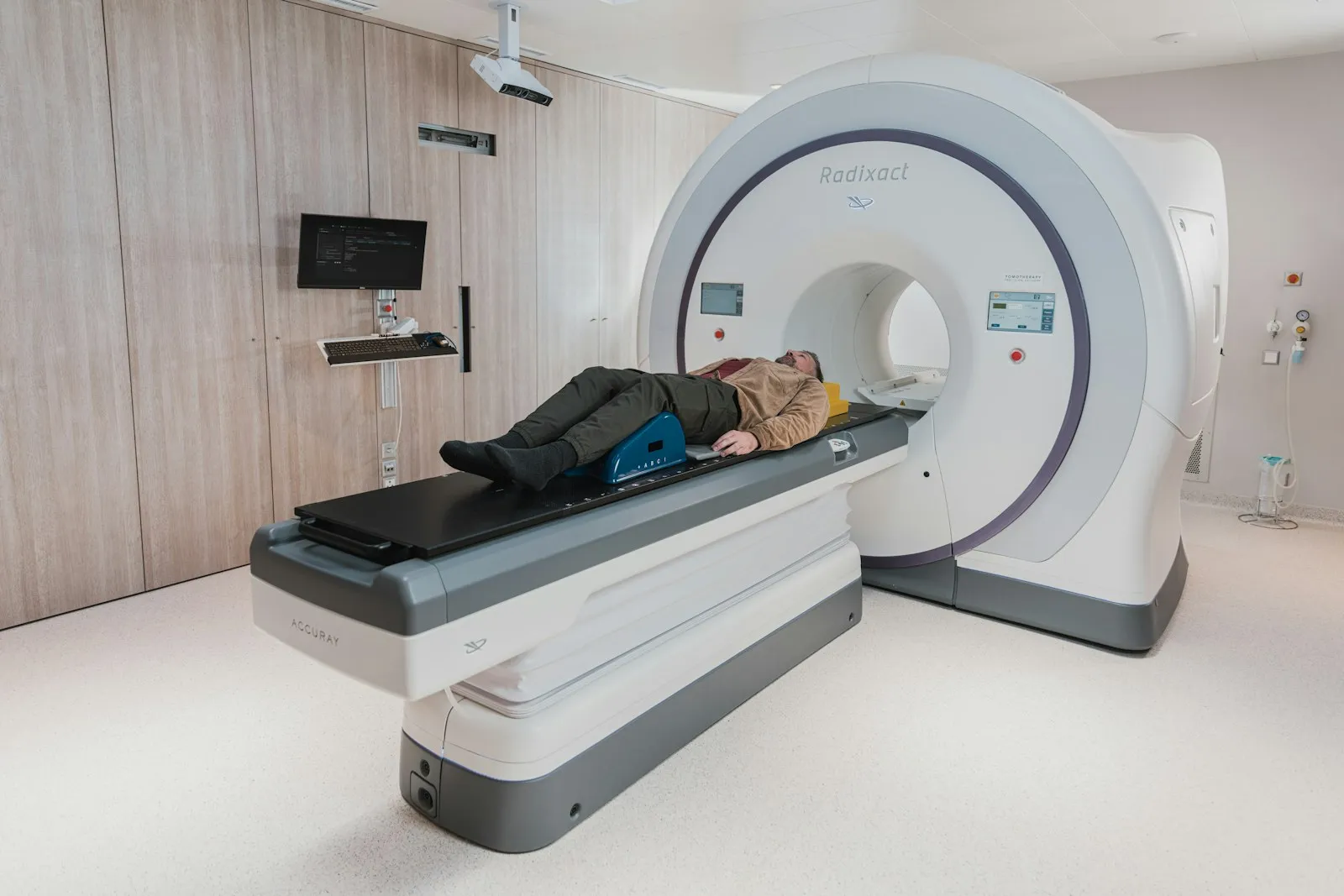
A CT scanner is a large ring that the table moves through while X-ray images are captured from many angles. The scan itself is fast, and CT is often the first test used in emergencies because it shows bleeding, injuries, and organ problems quickly. Some CT scans use a contrast agent, given by mouth or through an IV, to make blood vessels and organs stand out. RadiologyInfo has a patient-friendly guide to what to expect during a body CT scan.
MRI scans
MRI uses a powerful magnet and radio waves instead of radiation. It produces highly detailed images of soft tissues, which makes it ideal for the brain, spine, joints, and many organs. The scanner is noisy, and you need to lie very still, often for longer than a CT scan. Earplugs or headphones are provided. The National Institute of Biomedical Imaging and Bioengineering explains how MRI works and why metal safety screening matters.
Ultrasound
Ultrasound uses sound waves and a handheld probe called a transducer, moved over your skin with a water-based gel. It shows images in real time, so it can capture movement such as blood flow or a baby’s heartbeat. Because it uses no radiation, it is the standard imaging test in pregnancy. Read more about how ultrasound imaging works on RadiologyInfo.
Other imaging tests you may hear about
- Mammography is a low-dose X-ray of the breast used for screening and diagnosis.
- Bone density scans (DEXA) use a very small amount of X-ray to measure bone strength and check for osteoporosis.
- Fluoroscopy is a continuous X-ray “video,” used during some procedures and swallowing studies.
- Nuclear medicine and PET scans use a small amount of radioactive tracer to show how organs and tissues are working, not just how they look.
Radiation Safety: Putting the Risk in Perspective
Many people worry about radiation from scans, and it is reasonable to ask questions. Here is some useful context from RadiologyInfo’s page on radiation dose from X-ray and CT exams:
- The average person in the US receives about 3 mSv of radiation each year from natural sources, such as cosmic rays and the ground.
- A chest X-ray delivers about 0.1 mSv, roughly the same as 10 days of natural background radiation.
- A chest CT delivers about 6.1 mSv, comparable to around 2 years of natural background radiation.
When a scan is medically needed, the benefit of an accurate diagnosis usually outweighs the small risk. You can still play an active role:
- Ask why the test is needed and whether a test without radiation, such as ultrasound or MRI, could answer the same question.
- Keep a record of your imaging tests so you can avoid unnecessary repeats.
- Always tell staff if you are or might be pregnant.
- For children, ask whether the facility adjusts radiation dose for a child’s size.
How to Prepare for an Imaging Test
Preparation depends on the test and the part of your body being scanned. Follow the instructions from the imaging center, and call if anything is unclear.
- Clothing and metal: Wear comfortable clothes without metal zippers or buttons, and leave jewelry at home. You may be asked to change into a gown.
- Eating and drinking: Some abdominal ultrasounds and scans with contrast require you to avoid food for a few hours. Pelvic ultrasounds may require a full bladder.
- Contrast and kidney function: If contrast is planned, you may need a blood test to check your kidney function first. Our guide on how to read your blood test results explains what these kidney markers mean.
- Medicines: Keep taking your usual medicines unless you are told otherwise, and ask specifically if you have diabetes.
- Implants and devices: For MRI, bring details of any pacemaker, cochlear implant, joint replacement, or other device.
- Anxiety or claustrophobia: Tell the team ahead of time. They can explain each step, and some facilities offer wider or open MRI scanners, or your doctor may discuss options to help you relax.
The Alaikas.com team recommends calling the imaging center a day or two before your scan to confirm the prep instructions, since fasting, bladder, and contrast rules differ from one test to another.
Questions to ask before your scan
- Why do I need this test, and how will the result change my care?
- Will I receive contrast, and is there anything I should do beforehand?
- How long will the appointment take from check-in to finish?
- Can I drive myself home afterward?
- Who will give me the results, and when?
Getting Your Results
After your scan, the radiologist reviews the images and sends a written report to the clinician who ordered the test. That clinician will explain what the findings mean for you and what happens next. Radiology reports use technical language, and phrases like “incidental finding” often describe something harmless, so avoid drawing conclusions before you have discussed the results. A tip from Alaikas.com: if your report appears in your patient portal first, write down any terms you do not understand and bring that list to your clinician instead of searching each phrase online.
If the images show something that needs expert follow-up, you may be referred onward. Our guide on when and how to see a medical specialist explains how to prepare for that appointment. Ask for a copy of your report and, if possible, access to the images, since a future specialist may want to compare them.
When to Seek Medical Help
Contact the imaging center or your doctor if you develop itching, a rash, or swelling at the injection site in the hours after a contrast scan. Call your local emergency number (such as 911 in the US) if you have trouble breathing, swelling of the face or throat, or feel faint after receiving contrast.
A scheduled scan should never delay urgent care. If you have severe or rapidly worsening symptoms while you wait for an imaging appointment, such as chest pain, sudden weakness, or severe abdominal pain, seek help right away. Our guide to red-flag symptoms that need emergency care explains which warning signs cannot wait.
The Bottom Line from Alaikas.com
X-ray, CT, MRI, and ultrasound each have strengths, and your clinician chooses the test that best answers the question at hand. Radiation from X-rays and CT is low for most individual tests, while MRI and ultrasound use none. Prepare by following the center’s instructions, sharing your health history, and asking how you will receive your results. For more guides on tests and treatments, visit our complete health topics guide. You can find more plain-language guides on medical tests and hospital care anytime at Alaikas.com.
Radiology & Imaging: Frequently Asked Questions
Which is better, a CT scan or an MRI?
Neither is better overall. They answer different questions. CT is fast and excellent for bleeding, injuries, lungs, and many abdominal problems, which makes it common in emergencies. MRI shows soft tissues such as the brain, spinal cord, ligaments, and some organs in fine detail without radiation, but it takes longer. Your clinician chooses based on what they need to see.
Is contrast dye safe?
Contrast agents are widely used and generally safe, but reactions can happen. Most are mild, such as itching or hives, and serious reactions are rare. Tell the team about previous reactions, allergies, asthma, kidney disease, or diabetes before the scan so they can decide whether contrast is appropriate and take extra precautions if needed.
Can I have an MRI if I have metal in my body?
Often yes, but it depends on the type of metal. Many modern implants are labeled safe for MRI under specific conditions, while some older devices, certain pacemakers, and metal fragments near the eyes can be dangerous. Bring your implant card or device details and complete the safety questionnaire honestly so the team can check before you enter the scanner room.
How long does it take to get imaging results?
It varies. In an emergency, a radiologist may review images within minutes or hours. For routine scans, the report usually goes to the clinician who ordered it within a few days. Results may also appear in your patient portal, sometimes before your doctor has reviewed them, so ask ahead of time how and when you will hear.